
Discovering an unexpected lump on your gums can be concerning, especially when trying to determine if it’s a gum cyst vs blood blister. Both conditions present as raised lesions in the mouth, but they have fundamentally different origins, appearances, and treatment requirements.
A blister with blood in the mouth typically appears suddenly following minor trauma and resolves within two weeks, while a gum cyst develops gradually over weeks to months and persists indefinitely without intervention. Understanding these distinctions helps you respond appropriately and know when professional dental care is necessary.
This comprehensive guide examines the key differences between gum cyst vs blood blister, including their causes, visual characteristics, symptoms, and treatment approaches. Whether you’ve noticed a blood blister inside your mouth or suspect a gum cyst, this expert analysis provides the information you need to make informed decisions about your oral health.
What Is a Gum Cyst? Understanding Dental Cysts
A gum cyst, also called a dental or periodontal cyst, is an epithelial-lined cavity containing fluid or semi-solid material that develops in or around the gums. Unlike a blood blister in the mouth that contains blood, gum cysts typically contain clear to yellowish fluid, inflammatory cells, or tissue debris.
Common types of gum cysts:
Periapical cysts develop at the root tip of a dead or infected tooth and represent the most common type, accounting for approximately 60 percent of all jaw cysts. These form when bacteria from tooth decay or trauma cause root canal infection, triggering an inflammatory response that creates a fluid-filled sac.
Lateral periodontal cysts occur along the side of a tooth root in the periodontal ligament space. These developmental cysts arise from remnants of dental tissue and typically affect the lower premolar and canine regions.
Dentigerous cysts surround the crown of an unerupted tooth, most commonly impacted wisdom teeth. These developmental cysts form during tooth formation and can grow large enough to cause jaw expansion if left untreated.
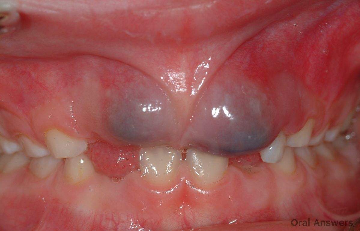
Eruption cysts appear as bluish swellings over erupting teeth in children, essentially representing dentigerous cysts that have reached the oral mucosa. These usually resolve spontaneously as the tooth breaks through.
Gum cysts develop gradually over weeks, months, or even years through chronic inflammatory processes or developmental abnormalities. The slow accumulation of fluid within the epithelial lining causes progressive expansion, unlike the sudden appearance of causes of oral blood blisters. Without intervention, gum cysts remain indefinitely and may continue enlarging, contrasting sharply with blood blister inside cheek formations that resolve naturally within 7 to 14 days.
What Is a Blood Blister in the Mouth?
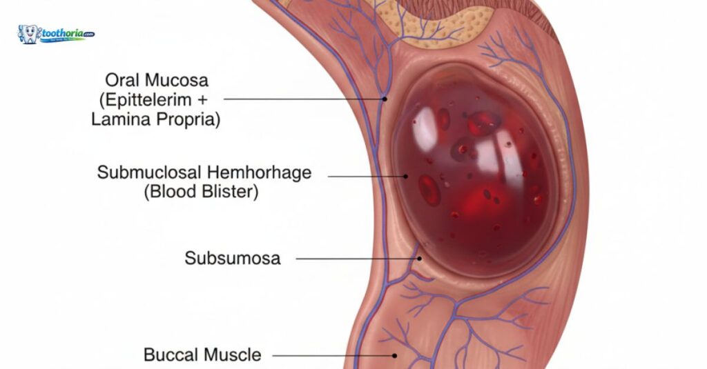
A blood blister in the mouth, medically termed “angina bullosa hemorrhagica,” is a subepithelial blister containing blood that forms suddenly on oral soft tissues. These benign lesions result from minor trauma or negative pressure that damages delicate capillaries beneath the mucosa.
Key characteristics:
Blood blisters appear within 1 to 4 hours of the triggering event, presenting as dark red, purple, or bluish-black raised lesions with smooth surfaces. They typically measure 3 to 30 mm in diameter and feel soft and fluid-filled when palpated. Most commonly affecting the soft palate (52 percent) and buccal mucosa (20 percent), these lesions can also develop on the tongue, lips, or rarely on the gums.
The natural evolution follows a predictable pattern: rapid formation reaching maximum size within 24 hours, spontaneous rupture within 24 to 48 hours releasing dark blood-tinged fluid, and complete healing within 7 to 14 days without scarring. This self-limiting nature fundamentally differs from the persistent characteristics of gum cysts, making it easier to distinguish gum cyst vs blood blister based on timeline alone.
Common causes of mouth blood blisters:
- Accidental cheek or lip biting during eating
- Nocturnal teeth grinding (bruxism)
- Aggressive tooth brushing
- Hard or crunchy food consumption
- Dental procedures involving suction
- Drinking through straws vigorously
- Nutritional deficiencies (vitamin C, iron)
- Medications affecting coagulation
Gum Cyst vs Blood Blister: Critical Comparison
Visual and Physical Differences
Understanding appearance differences between gum cyst vs blood blister enables accurate identification:
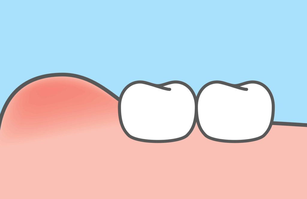
Gum cyst appearance: A gum cyst typically presents as a smooth, dome-shaped swelling on the gum tissue with normal pink coloration or a slight bluish tint. The overlying mucosa appears intact and stretched over the firm mass beneath. When palpated, cysts feel firm and resilient, similar to a small rubber ball, and do not compress or change shape with gentle pressure.
Blood blister appearance: In contrast, a blood blister inside the mouth displays distinctive dark coloration ranging from deep red to purple or nearly black. The lesion appears as a raised bubble with a tense, shiny surface that clearly contains fluid. Gentle palpation reveals soft, fluctuant consistency.
Diagnostic Comparison Table
| Feature | Gum Cyst | Blood Blister in Mouth |
| Onset | Gradual (weeks to months) | Sudden (1 to 4 hours) |
| Color | Skin-colored, slightly bluish | Dark red, purple, or black |
| Texture | Firm, non-fluctuant | Soft, fluid-filled |
| Contents | Clear fluid, debris | Blood |
| Location | Always near teeth/roots | Any oral mucosa |
| Duration | Persists indefinitely | Resolves in 7 to 14 days |
| Pain | Usually painless | Mild to moderate |
| X-ray | Visible as dark area | No radiographic changes |
| Treatment | Professional removal needed | Home care sufficient |
Symptoms and Clinical Presentation
Gum Cyst Symptoms
Most small to moderate gum cysts remain asymptomatic and are discovered during routine dental examinations or radiographs. The firm, painless swelling on the gum near a tooth may be noticed by the patient or dentist during visual inspection.
When symptoms occur:
- Visible facial asymmetry or jaw expansion
- Throbbing pain if cyst becomes infected
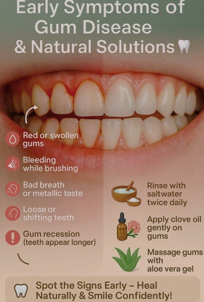
- Gum redness and swelling
- Abscess formation with pus drainage
- Tooth mobility when cysts expand around roots
- Difficulty with denture fitting
Blood Blister Symptoms
A blood blister on the tongue or other oral location produces distinct symptoms:
Initial phase:
- Sudden awareness of oral mass or bubble
- Dull aching or pressure sensation
- Sharp pain when compressed by teeth
- Metallic or blood taste
After rupture:
- Brief sharp pain upon rupture
- Release of dark blood-tinged fluid
- Burning or stinging sensation
- Increased sensitivity to hot, cold, or acidic foods
These symptom patterns help clinicians and patients differentiate gum cyst vs blood blister presentations.
Diagnosis and Professional Evaluation
Dental professionals use systematic approaches to differentiate these conditions:
Clinical examination steps:
Visual inspection assesses lesion location, size, color, and relationship to teeth. Gum cysts demonstrate consistent association with specific teeth, while inside-mouth blood blister formations can occur anywhere on the oral mucosa without dental correlation.
Palpation testing clearly distinguishes firm, non-compressible gum cysts from soft, fluctuant blood blisters. Mobility testing reveals that cysts remain fixed to underlying bone or tooth structures, while blood blisters move freely with surrounding soft tissue.
Radiographic evaluation provides a definitive diagnosis for gum cysts, showing characteristic radiolucent (dark) areas around tooth roots or within the jawbone. Blood blisters produce no radiographic changes since they involve only soft tissues without bone involvement.
When to seek professional care:
- Any firm gum swelling persisting beyond two weeks
- Lesions associated with tooth pain or sensitivity
- Facial swelling or asymmetry
- Signs of infection (pus, fever, severe pain)
- Blood blisters recurring frequently (3 or more in 6 months)
Treatment Approaches
Gum Cyst Treatment
Professional intervention is mandatory for gum cysts:
Root canal therapy: When periapical cysts result from tooth infection, successful root canal treatment often allows small cysts to resolve naturally as infection clears. The procedure removes infected pulp tissue, disinfects root canals, and seals the system to prevent bacterial reentry.
Apicoectomy: For cysts persisting after root canal treatment, surgical removal of the root tip and cyst becomes necessary. The procedure involves accessing the root through the bone, removing the cyst lining, and sealing the root end.
Enucleation: Complete surgical removal of the cyst lining ensures definitive treatment and prevents recurrence. The dentist or oral surgeon removes the entire cyst capsule and may place bone graft material in large defects.
Tooth extraction: When cysts involve severely damaged teeth beyond restoration, extraction of the affected tooth along with cyst removal provides the most practical solution.
Blood Blister Treatment
Conservative home management suffices for blood blisters in the mouth:
Immediate care steps:
- Avoid intentional rupture or manipulation
- Rinse gently with cool water
- Apply ice externally to reduce swelling
- Document with photos for tracking
Ongoing management:
- Saltwater rinses 4 to 6 times daily (one-half teaspoon salt in 8 ounces warm water)
- Soft diet avoiding rough, hot, or acidic foods
- Topical anesthetics containing benzocaine for comfort
- Over-the-counter pain relief (ibuprofen 400mg every 6 to 8 hours)
Professional intervention is rarely needed except for exceptionally large lesions causing functional impairment or recurrent cases requiring underlying cause investigation.
Frequently Asked Questions About Gum Cyst vs Blood Blister
Why does my gum have a blood blister?
Gums rarely develop true blood blisters since they consist of keratinized tissue. What appears as a blood blister on gums may be an eruption cyst in children, a vascular lesion, or a gum cyst. Professional evaluation provides accurate diagnosis.
How do I know if I have a cyst in my gum?
A gum cyst appears as a firm, painless swelling near a tooth that persists for weeks without healing. It feels hard when pressed and shows as a dark area on dental X-rays. Professional diagnosis requires clinical and radiographic examination.
What is a lump on a child’s gums?
The most common lump is an eruption cyst, a bluish fluid-filled swelling over an erupting tooth that resolves naturally. Other possibilities include dental abscesses from cavities or developmental cysts. Consult a pediatric dentist if the lump persists beyond two weeks.
What does an oral blood blister look like?
An oral blood blister appears as a raised, dome-shaped lesion with dark red, purple, or black coloration. The smooth surface looks like a bubble filled with dark fluid, typically measures 5 to 20 mm, and feels soft when touched.
Conclusion: Know When to Act
Understanding the differences between a gum cyst vs blood blister empowers you to respond appropriately to oral lumps and lesions. While a blood blister inside the mouth represents a benign, self-limiting condition requiring only conservative home care, a gum cyst indicates underlying dental pathology necessitating professional treatment to prevent complications.
The key distinction lies in onset speed, appearance, and duration. Blood mouth blisters appear suddenly with characteristic dark coloration and resolve within two weeks, while gum cysts develop gradually as firm, persistent swellings associated with specific teeth. When facing uncertainty about any oral lesion, especially firm gum swellings persisting beyond two weeks, professional dental evaluation provides definitive diagnosis and appropriate treatment.